Thursday, August 3, 2017

Painful neuromas - why they occur and what we can do about them

Painful neuromas - why they occur and what we can do about them

One of the most common nerve conditions that I treat is the painful neuroma. These are some of the most challenging issues to treat, requiring a very careful eye for diagnosis AND a thoughtful approach to treatment.

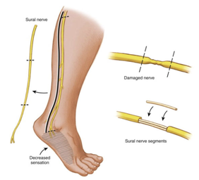
Normally, nerves are smooth and flowing structures (Figure A). Neuromas form after a nerve has been cut/injured* (Figure B) - by default, the body's default response to this is the prepare the near-end of the nerve to regenerate: it automatically tries to find the far-end of the nerve. If the environment won't allow the near-end to find the far-end (either because the gap is too much to overcome or due to lots of scarring around the injury), nerve regeneration will not occur (Figure C). The near-end is constantly seeking the far-end and remains in an inflammatory state, leaving it very irritable and causing pain. The near-end of the nerve can be rubbed on by the moving skin and fascia, creating remarkable sensitivity that is unbearable. This is the type of pain that patients often describe as sharp, shooting, shearing, "ice pick" or stabbing, starting at the area of nerve injury and heading/radiating to somewhere further out on the arm/hand or leg/foot.

*"neuromas-in-continuity" can appear with long-standing compression of a nerve or with scarring of a repaired nerve


Now we are left with a tough situation in which a nerve is trying to regenerate but is instead causing pain. So what can we do about it? Most nerve surgeons will provide the response that the best treatment is prevention of nerve injuries. YES, THIS IS ABSOLUTELY TRUE... but not helpful for the patient who is already suffering from a nerve injury.

For those patients with painful neuromas, we can try a few different options. In my experience, if we can isolate the patient's symptoms to a specific portion of the nerve that is inflamed, we can target our treatment to this area. I typically send my patients for ultrasound-guided injections to the suspected painful neuroma region, and if this provides even temporary and/or partial relief, this is a good sign that I can help the patient with surgery. If I am having a hard time localizing the patient's symptoms to one specific nerve, I will typically start with medications for nerve pain (such as gabapentin, pregabalin, nortryptyline, amitryptyline, or duloxetine).

For those patients who respond well (or even somewhat well) to an injection, I recommend surgical treatment of the neuroma. The near-end of the nerve is trying to regenerate, so I prefer to target our treatments to help it find the far-end. In surgery, we freshen the near and far ends of the nerve, then place a nerve graft (sometimes autograft, such as from the sural nerve... and sometimes allograft from a cadaver) to lay the "train tracks" for the nerve to regenerate  (Figure D). If I cannot do that reliably, I will take the near-end of the nerve and bury it in muscle - while the nerve will not regenerate, it will at least find a "dead-end" in the muscle, where it will not be irritated by constant contact from the skin (Figure E). In both of these situations, my goal is to RELIEVE PAIN... and I counsel all patients that they are almost certainly trading off their pain but getting some numbness in return.

Painful neuromas are a very challenging problem, but I have also found patients with this issue to be some of the most grateful patients I have. Careful diagnosis, thoughtful treatment, and appropriate counseling about expectations are the key to helping patients with this condition.


Christopher J. Dy, MD MPH
My Bio at Washington University Orthopedics
dyc@wustl.edu 


Friday, June 30, 2017

Sural nerve harvest - no big deal, right?

Sural nerve harvest - no big deal, right?

Image result for cabled graft nerveI wanted to write something about sural nerve harvest because it is something I discuss with patients nearly every time we are talking about treatment of nerve injuries. We will often use the sural nerve as an autograft during nerve reconstruction cases. After we remove the scarred/injured part of the nerve, there is usually a gap that we cannot repair directly. We will suture the sural nerve graft to bridge the gap between the nerve ends, often laying down multiple segments of the graft ("cabling" the graft) to replicate the thickness of the nerve we are replacing.

How is the sural nerve harvested? The nerve is identified through either one long incision or a series of small incisions along the back of the calf (just behind the smaller bone in the shin - the fibula). The nerve is identified near the ankle level and traced up to the knee level. After the sural nerve is cleared from the surrounding tissue, the nerve is cut, removed from the body, and prepared for grafting.

What are the downsides of harvesting the sural nerve? Like they say, there's no free lunch.
Image result for sural nerve
  • Numbness: The sural nerve's normal function is to provide sensation to the back of the calf and the outer border of the foot (area seen in purple). Removing the sural nerve will lead to numbness in this area, which is most noticed when walking barefoot or in sandals, or when playing sports like soccer/football that require contact with the outside of the foot. All patients will almost certainly experience this. 
  • Pain: Anytime a nerve is cut (including for a harvest), the nerve ends are very sensitive since they are trying to regenerate. Most of the time, the near-end of the nerve retracts into the calf muscles and doesn't see much irritation since it is buried so deeply. But in some patients (about 5%), the nerve can be very sensitive (painful neuroma). Most of the time, this will get better over time (about 3-6 months). Medications for nerve pain may be helpful. Very rarely, another surgery is needed to re-cut the nerve ends and bury them deep in muscle or bone.
  • Infection, hematoma, wound healing problems, and blood clots (deep venous thrombosis) - these can happen in any leg surgery and certainly need to be mentioned. 
Does the numbeness ever get better? Over time, the numbness will eventually decrease as the remaining nerves grow into the area that the sural nerve used to supply. It will never feel "normal", but eventually will lead to the ability to tell the difference between hot and cold (what we call protective sensation)... except in kids, who often times cannot tell a difference because their nerves grow so quickly.

Are there any other options? Nerve grafts can be harvested from other sources (such as the saphenous nerve in the thigh, the medial antebrachial cutaneous nerve around the elbow, and the posterior interosseous nerve in the wrist) instead of (or in addition to) the sural nerve. The sural nerve tends to be many surgeons' preferred choice (including mine) because it has the highest percentage of nerve fibers (fascicles), a lot of graft can be harvested (up to 35-40cm), and the downsides seem to be tolerated well. Cadaver nerve graft (allograft) is another option, but the studies are still being done to show if it is "good enough" when compared to using the patient's own nerve (autograft). One of the biggest issues with the allograft is that the process to make the allograft non-reactive and non-infectious removes some of the cells that promote nerve regeneration (Schwann cells).


Christopher J. Dy, MD MPH
My Bio at Washington University Orthopedics
dyc@wudosis.wustl.edu 

Tuesday, June 20, 2017

Peroneal nerve injuries - one tough cookie!

While the majority of my practice is in the upper extremity, one of the things I enjoy the most about specializing in nerve surgery is getting to team up with colleagues from all of the different orthopedic/plastic subspecialties. Several times a month, I see patients with nerve injuries in their legs - sometimes with compressive neuropathies like tarsal tunnel syndrome, or those who have had nerve injuries from motor vehicle accidents or falls. 

One of the more common lower extremity nerve injuries that I see involves the peroneal nerve. I wanted to spend some time writing about these injuries to help out any of my patients (or anybody else out there) seeking more information about them.
Image result for aaos peroneal nerve
What is the peroneal nerve? The common peroneal nerve is a branch of the sciatic nerve that "comes into its own" at the lower part of the thigh, just above the knee; it then continues on the outside (lateral) part of the knee and wraps around the neck of the fibula bone (the skinny bone on the outside of the shin). The common peroneal nerve is "common" because it has 2 of its own branches - the deep branch and superficial branch. Both of these nerve branches have motor and sensory jobs. The deep peroneal nerve supplies the very important muscles that allow you to raise your ankle (tibialis anterior) and toes (extensor hallucis longus and extensor digitorum longus), but also carries the sensation from the space between your big toe and second toe. The superficial peroneal nerve supplies the muscles that swing your ankle outside (eversion by the peroneus longus and brevis muscles) and carries sensation from the top part of your foot (closer to the second through little toes).

How does a peroneal nerve injury occur? The peroneal nerve gets stretched during injuries that place stress on the outside of the knee - such as dislocations of the knee, ligamentous damage to the knee (such as the ACL or LCL), or fractures of the femur, tibia or fibula. These injuries can happen after falls or motor vehicle accidents, or from many other causes. As the peroneal nerve is stretched during the injury, the inner portions of the nerve may stretch and snap/rupture. In severe injuries, the entire nerve may rupture. Scar tissue forms around the injured parts of the nerve and the nerve can no longer conduct the motor and sensory functions. 

What is the prognosis? Recovery after any nerve injury (upper or lower extremity) is tough - it takes a long time for recovery and the function/strength is never "quite the same" as before the iinjury. Unfortunately, peroneal nerve injuries are among the toughest to treat - there tends to be a lot of scarring aroud the nerve ends as the very strong calf muscles pull the nerve ends apart. Additionally, the internal composition of the peroneal nerve isn't as favorable for a fast recovery. Despite advances in microsurgery, the results from nerve repair, nerve grafting, and nerve transfers have not been as successful for the peroneal nerve as nerves in the shoulder/arm (brachial plexus). 

What are the treatment options? If the peroneal nerve injury is diagnosed early enough, your surgeon may choose to explore the peroneal nerve and remove scar tissue around/within the nerve (neurolysis). If there is a gap between the nerve ends, the surgeon may choose to place a nerve graft to provide a scaffold for nerve regeneration. If the gap is too big for a chance of recovery with a graft, one option is to perfrom a nerve transfer, in which an expendable working nerve (to either curl the toes or press the ankle down) is connected to the muscles supplied by the peroneal nerve. There are two big issues with nerve transfers for peroneal nerve palsies: it's really hard to re-train the brain to make the nerve transfer work and the results from these transfers have not been reliably good. For this reason, your surgeon may discuss tendon transfers with you - in this procedure, working tendons are re-routed to help bring the ankle and toes up. This is the most reliable "tried and true" procedure, and is often what we use for patients with this tough problem. It doesn't help with sensation to the top of the foot. One of the best parts about working at Washington University Orthopedics is that I get to team up with some of the best foot and ankle surgeons in the country - we work together to get patients the best outcome possible.  

Overall, this is a very challenging injury. I am very honest with my patients about the tough road ahead of them. I will try my best to restore the innervation to the muscles and sensation of the foot, but tendon transfers may be necessary in the future. Seeing eye-to-eye on expectations and goals for functional recovery is important for all nerve injuries, but especially for peroneal nerve injuries. 

Christopher J. Dy, MD MPH
My Bio at Washington University Orthopedics
dyc@wudosis.wustl.edu 

Wednesday, February 1, 2017

Nonoperative Treatment of Brachial Plexus Injuries

The last blog post talked about a list of brachial plexus injury topics that patients and their families want more information about... so let's start with talking about nonoperative treatment for brachial plexus injuries. 

One of the most important things to me is seeing patients with suspected brachial plexus injuries relatively soon after their injury, ideally within the first 6 weeks. This lets me establish a relationship with my patients long before we "need" to talk about surgery, since there is often a good chance that the nerve injury will recover without surgery (although lots of time, patience, and therapy is needed to get back to function). 

When I see a patient early on after their injury, we talk about the "basics" - what the brachial plexus is, how the nerves work, how they are usually stretched but can sometimes be pulled out of the spinal cord, and that it takes about 3 months to start to see signs of recovery (not full recovery... but hints that things are going to improve over time). During this initial 3-4 months after their injury, we focus on physical therapy to strengthen the remaining muscles (especially those around the shoulder blade/scapula), stretching to keep the joints supple, getting any nerve-related (neuropathic) pain under control, allowing other injuries (like fractures/broken bones) to mend, and (very importantly) keeping a healthy mind state (since depression and anxiety come up more than we typically expect). 

We usually get a nerve test (EMG) at the 6 week mark and at the 3 month mark, allowing me to compare whether the muscle recovery has progressed over time. Usually at 3 months, we'll see some slight hints of recovery and we can make some decisions about whether to proceed to surgery (if there isn't any recovery) or whether to wait and see what recovers (if there are signs of muscle activity). If we wait and see, we'll get another EMG and I'll examine the patient again in about 6 weeks. If things are moving in the right direction, we'll let the recovery mature and check how things are in another 1-2 months. If there aren't any signs of muscle recovery, we'll discuss surgical options again.

Establishing an early relationship with my nerve injury patients is important to me, since I often end up being the "quarterback" or captain of their care - long after their other injuries have healed, treatment for the nerve injury is front and center. I would rather see patients early on and watch them heal without surgery than meet patients for the first time a little later than I prefer... and have to tell them that they should have surgery.

Just a couple of thoughts for now - I'll follow this up with a brief Q&A with a patient who went through the nonoperative treatment process with me... and is doing incredibly well!